
Detailed ultrasound during pregnancy
Thanks to detailed ultrasound technology, vital information about baby and maternal health can be collected. Such imaging methods are very valuable for early detection, especially in the early stages of pregnancy, for detecting any abnormalities or health problems that may pose a risk to the baby in the future.
These imaging methods are also important in later gestation periods, helping to determine the criteria used to decide the baby’s blood flow, general development, health status, and type of delivery (normal or cesarean section).
Such high-detail ultrasound examinations are usually performed by perinatology specialists and the results are evaluated within the scope of High-Risk Pregnancies and Perinatology.
Detailed ultrasound during pregnancy
“Detailed Ultrasound” or “Detailed Ultrasound” is performed with a higher image quality than standard ultrasound examinations. In this special examination, more complex details about the baby and organs are examined in detail.
These examinations are critical not only for the health of the baby and mother, but also for future planning and treatments.
When is a detailed ultrasound done during pregnancy?
- Between 11-13 weeks: Suitable for all pregnancies.
- Between 18-23 weeks: Again, it is recommended for all pregnancies.
- Week 24 and later: Recommended for high-risk pregnancies.
Such examinations are usually carried out by perinatologists who have experience in congenital disorders and diseases. Ultrasound technology provides detailed information without harming the mother and baby.
The detailed ultrasound report provides detailed anatomical information about the current health status of the baby and the mother. In addition, if necessary, this report can also be a guide for future steps and counseling.
If you want to know more, you can scroll down or ask additional questions. Such examinations are critical for maternal and infant health, as well as for future medical decisions.
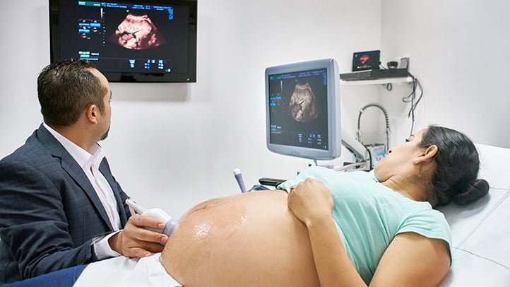
First Trimester Ultrasound in Early Pregnancy
Ultrasonography performed in the early stages of pregnancy, that is, within the first three months when the embryo is placed in the mother’s womb, provides a lot of important information and is vital for detecting potential risks early. This procedure is not harmful to either the mother or the baby.
The report covers the following topics:
- The number of gestational sacs and their location in the uterus
- Determining whether the pregnancy is ectopic (ectopic pregnancy)
- Evaluation of the anatomical structure and development of the gestational sac
- Measurement of the rate and rhythm of the baby’s heartbeat
- In twin or multiple pregnancies, determining whether the partners of the babies are the same or different
- Examination of the condition of the cervix
- Whether there is any bleeding that may be associated with the gestational sac
Especially in health centers specializing in High-Risk Pregnancies and Perinatology, patients are subjected to this early ultrasonography and the results are presented in a detailed report. This report may contain critical information for future medical interventions and treatment plans.
11-13th Week Detailed Ultrasound Examination
This period is the time period when the first comprehensive ultrasonographic evaluation is performed for the developing baby in the mother’s womb. This examination, which is carried out both from the abdomen and vaginally, is considered the first stage of a detailed ultrasonographic evaluation of pregnancy.
The information obtained from this review is as follows:
- Determination of the baby’s developmental week
- Location of the placenta (baby’s partner)
- The amount of amniotic fluid (baby’s water)
- Measurement of the nuchal translucency of the baby
- Examination of the baby’s nasal bone
- Basic observations on the baby’s heart structure
- Initial information about other areas of development of the baby, such as the brain, stomach, kidneys, bladder, abdominal wall, intestines, spine, hands and feet
- Examination of the specific vascular structures of the baby using color Doppler
- Assessment of the condition of the cervix
As a result of this examination, all these data obtained and your doctor’s observations are presented to you in a detailed report. This first comprehensive ultrasound examination allows you to assess your baby’s genetic and morphological (seen) abnormalities for the first time. Especially in health centers specialized in the field of High-Risk Pregnancies and Perinatology, this 11th-13th week ultrasonography is routinely applied.
18-23 Week Detailed Ultrasound Review
After a detailed ultrasonography examination in the first trimester, a second comprehensive ultrasonographic evaluation is performed at 18-23 weeks of pregnancy.
This second detailed ultrasonography is one of the critical elements of pregnancy follow-up. In addition to the data obtained by the initial examination, a more detailed assessment of the organs and anatomical structure of the baby is usually carried out during this period. In addition, since the baby grows in this process, it is an ideal time period for the detection of more complex structures and possible abnormalities.
The examinations usually performed during this period provide more comprehensive and detailed information about many organs and systems of the baby such as heart structure, brain, kidneys, spine, extremity development.
Detailed ultrasonography between the 18th and 23rd weeks is routinely applied in health centers specialized in High-Risk Pregnancies and Perinatology, and the results of this examination are very important for the health of the mother and baby. The data obtained becomes a valuable guide when planning the birth and performing medical interventions when necessary.
How is Detailed Ultrasonography Performed?
Below you will find a step-by-step explanation of how the detailed ultrasonography procedure is performed:
Making an Appointment:
The procedure can usually be applied from the 18th week of pregnancy. Families can make an appointment for a date that suits them by calculating which week of pregnancy it is (usually between 18-23 weeks).
Appointment Day:
The process usually takes 30-45 minutes, but this period can sometimes take up to 1 hour or may need to be repeated in the following days.
Should you go to a detailed ultrasound hungry or full?
You do not need to be hungry for the procedure, you can maintain your normal diet.
How to do a detailed ultrasound:
This is usually an abdominal ultrasound procedure. After the procedure, the condition of the cervix is also evaluated and, if necessary, additional ultrasonographic examinations are performed vaginally.
Report Content:
The detailed ultrasonography report contains detailed information about the current status of the pregnancy and the anatomy of the baby. The report provides extensive information about the baby’s health status at that time and also offers counseling suggestions when necessary.
These stages play a critical role, especially in the management of high-risk pregnancies and birth planning. Detailed ultrasonography is accepted as a standard practice in specialist health centers and the data obtained are very valuable for maternal and infant health.
Can Detailed Ultrasound Be Done a Second Time?
If you have already performed this procedure in a different health center and are considering doing it again, this is possible. Ultrasonography is not harmful to you or your baby.
Can It Be Done Again in Advanced Pregnancy Weeks? Although detailed ultrasonography is generally recommended between 18-23 weeks, this type of examination can be repeated in the following weeks of pregnancy for special cases and re-evaluation of certain regions.
In the field of Perinatology and High-Risk Pregnancy, detailed ultrasonographic examinations are performed. Especially in high-risk pregnancies, such detailed examinations can be critical and provide valuable information for the health of the mother and baby.
Fetal Heart Ultrasonography – Echocardiography
Fetal echocardiography is a special type of ultrasound in which the heart of the baby in the womb is examined in detail. It is usually performed between 18-23 weeks, but it can also be done at different times depending on special conditions and risk factors.
Cause 22. Week Important? The 22nd week is a period when the fetal heart can be evaluated in more detail. Some heart diseases can be seen more clearly in this week.
Who is it recommended for? Fetal echocardiography is usually recommended for pregnancies with the following risk factors:
- Families with babies with heart anomalies in previous pregnancies
- Expectant mothers with risk factors such as metabolic diseases (e.g., diabetes)
- Families with congenital heart defects in the expectant mother or father
- Twin pregnancies with identical twins
- Expectant mothers with epilepsy
- Expectant mothers who have some infections in the early stages of pregnancy
- Expectant mothers using certain medications
- Rounds 1 and 2 Infants whose suspicion of fetal heart disease could not be excluded in trimester ultrasonography
This type of examination provides a more detailed heart evaluation in pregnancies with the above risk factors or suspicious conditions. In addition, such examinations can often be repeated in the last trimester of pregnancy, especially if possible heart problems have been detected.
In the field of Perinatology and High-Risk Pregnancy, detailed fetal echocardiographic examinations are provided. These examinations offer important information that can be critical to the health of the mother and baby.
Detailed ultrasounds after the 24th week
In the standard process of pregnancy, detailed ultrasonography procedures are performed in certain weeks. However, sometimes detailed ultrasonographic examinations can be performed in advanced gestational weeks. This usually applies to special cases such as:
- The appearance of certain health problems
- Detection of abnormalities in previous ultrasonography results
- The occurrence of a risk factor in the mother or baby
- The patient misses the planned ultrasonography date for some special reasons
In Perinatology and High-Risk Pregnancies, detailed ultrasonographic examination is also performed on patients admitted at 24 weeks and later. This is especially important to identify complications or risks that occur in the late period.
What kind of details are examined in detailed ultrasonography?
- Baby’s developmental week
- Organs and anatomical structure of the baby
- Movements and position of the baby
- Baby’s heart rhythm
- The amount of fluid in the baby
- Location and condition of the placenta
- Evaluation of the cervix
Such detailed examinations can be critical to assessing potential risks to both mother and baby and providing an opportunity for early intervention. The results of the examinations are given to the patient in the form of a report and a follow-up plan is created when necessary.
Color Doppler Review
Color Doppler ultrasound visually shows the flow of blood through the vessels, and such examinations are usually performed more often in high-risk pregnancies. It is especially used to evaluate the vascular structure of the baby and the placenta. Color Doppler examination may be important for specific situations such as:
- Conditions with growth retardation
- Decrease in the baby’s amniotic fluid
- Disruption of the baby’s circulation
- Evaluation of the baby’s heart function
- Decreased movement of the baby in the womb
- Features of placental blood flow of fetuses in multiple pregnancies
For such cases, Color Doppler can provide doctors with more detailed information, helping them make appropriate treatment or intervention plans.
Biophysical Profile Review (BPP)
The Biophysical Profile Review consists of a series of tests and observations of how the baby is in the womb. It is usually combined with an NST (Non-Stress Test) and gives detailed information about the baby’s health. The biophysical profile can evaluate the following parameters:
- Baby’s tone (the ability to contract and relax muscles)
- Baby’s respiration
- Amount of amniotic fluid
- Baby’s movements
These reviews offer valuable information about the baby’s overall health and well-being in the womb and are summarized through a scoring system. High or low scores can be a signal for additional investigations or interventions.
Both Color Doppler and Biophysical Profile Examination are invaluable tools for painting a comprehensive picture of the health of the mother and baby, especially in high-risk pregnancies. With such examinations, doctors can diagnose possible complications early and make appropriate treatment plans.